harvardapparatus.com is a data controller for the purposes of the General Data Protection Regulation ("GDPR"). By proceeding to tick the consent box at right, you consent to harvardbioscience.com processing your personal data, under the GDPR and any other applicable legislation, that we collect from you in accordance with our Privacy Policy. If you wish to withdraw your consent for us to process your personal data please contact our Compliance Team. Please address any questions, comments and requests (including your right to withdraw your consent to process your personal data) regarding our data processing practices to our Compliance Team.
Allow

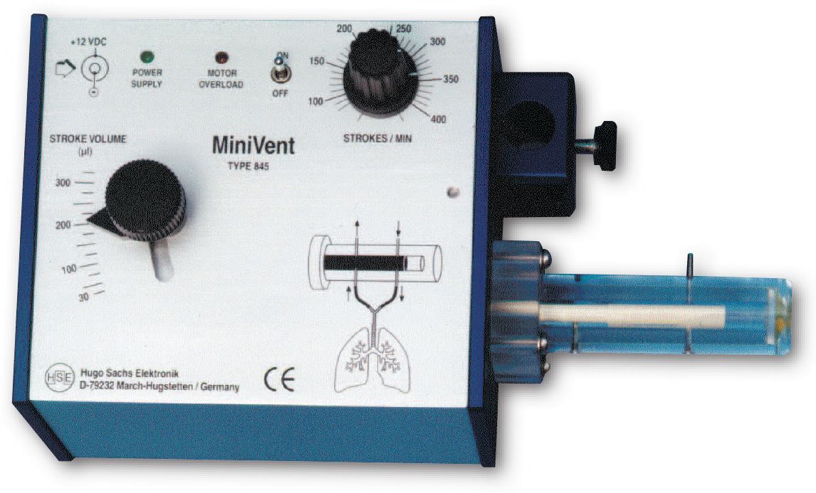
MiniVent Ventilator for Mice (Model 845), Single Animal, Volume Controlled

SKU
65509d00

The MiniVent Model 845 Ventilator is a quiet, compact and light weight ventilator. While it was designed specifically for mice, the MiniVent can be used for any animal (e.g. birds and perinatal rats) which requires tidal volumes in the range of 30 to 350 µl and respiratory rates of 60 to 400 breaths per minute.

  • Ideal ventilator for mice 
  • Stroke volume range from 30 to 350 µl 
  • Ventilation rate from 60 to 400 breaths/minute 
  • Simple adjustment of stroke volume while running 
  • Valveless piston pump, no valves to clog 
  • Very small instrument/circuit dead space volumne 
  • Compact construction, easy to install close to animal 
  • No vibrations, very low noise 

See our support center page here for information regarding replacement power supplies.


Grouped product items
Item No. Description Qty
73-0044 MINIVENT TYPE 845, 110V/220V; Tidal Volume 30-350 µl Please login to add to Quote
73-3077 ANESTHESIA MACHINE CONNECTION KIT FOR MICROVENT, MINIVENT AND MIDIVENT VENTILATOR. Please login to add to Quote
73-4472 PEEP System with Tube Set to provide positive expiratory pressure on a MiniVent, MicroVent or MidiVent Please login to add to Quote
73-2919 Multi-Gas Inlet Adapter and stand to connect Aerosol Nebulizer and MicroVent, MiniVent or MidiVent Please login to add to Quote
73-4013 Trigger Output Option for Minivent, End of Expiration Please login to add to Quote
73-4008 Trigger Output Option for MiniVent, End of Inspiration Please login to add to Quote

The MiniVent Model 845 Ventilator is a quiet, compact and light weight ventilator designed specifically for mice. It can be used for any animal (e.g. birds and perinatal rats) which requires tidal volumes in the range of 30 to 350 µl and respiratory rates of 60 to 400 breaths per minute.

Key Features

  • Ideal ventilator for mice 
  • Stroke volume range from 30 to 350 µl 
  • Ventilation rate from 60 to 400 breaths/minute 
  • Simple adjustment of stroke volume while running 
  • Valveless piston pump, no valves to clog 
  • Very small instrument/circuit dead space volumne 
  • Compact construction, easy to install close to animal 
  • No vibrations, very low noise


How it Works

The MiniVent Ventilator is a constant-volume respiration pump operating on the Starling principle. Unlike conventional units for larger animals, this ventilator employs a rotary plunger and has no valves. During each ventilation cycle, the plunger performs a synchronized forward and rotating movement. Cleverly arranged bores and channels in the cylinder and plunger control inspiration and expiration during each stroke of the plunger.

The extremely light weight and compact construction, in addition to the convenient rod clamp, allow the MiniVent ventilator to be positioned directly next to the animal. Typical setups with larger ventilators produce large tubing and instrument dead space volumes. These larger volumes introduce greater system compliance which can affect the accuracy with which the full tidal volume is introduced into the animal’s lungs. With the MiniVent, the tidal volume error due to system compliance is reduced to ±3 µl.

Tidal volume and respiration rate can be set exactly to the values required for mouse ventilation. The level of precision and control available to the investigator minimizes the danger of hyperventilation or hypoventilation.

The tidal volume can be varied continuously from 30 to 350 µl during operation without having to interrupt ventilation. The respiration rate is also continuously adjustable from 60 to 400 strokes/min. The expired air can be recovered at the collection port for sampling, recycling or for the generation of a positive end-expiratory pressure (PEEP). Room air or any non-explosive gas mixture can be used to feed the pump intake.


Components

The MiniVent is supplied with 1 x AC Wall Mounted Power Supply (115 V or 220 V); 2 x Silicone Tubing (1.5mm ID, 3.0mm OD, 14cm long); 1 x 1.3mm OD Tracheotomy Cannula (73-2730); 1 x 1.2mm OD Intubation Cannula (73-2844).

A multi-gas inlet adapter is available for the MiniVent so that alternate gas mixtures and nebulized substances are delivered to the MiniVent inlet port at atmospheric pressure. The adapter provides ports for multiple selectable gas mixtures (hypoxic, anesthetic...) and a port for the Aerosol Nebulizer.

Voltage Range
73-0043
73-0044

 115 VAC
 230 VAC
Number of Animals  1
Species  Mouse, prenatal rat, small bird
Control Modes  Volume
Gas Supply  Room air or non-flammable mixed gas
Display  None
IE Ratio,%  1:1
PEEP  Provided via attachment of water column
Respiration Rate  60 (min) to 400 (max) breaths/min
Respiration Rate Note  Continuously adjustable from 60 to 400 breaths/min
Sigh Frequency  None
Sigh Pressure  Not available
Tidal Volume  0.03 (min) to 0.13 (max) ml/stroke
Stroke Volume   Continuously adjustable from 30 to 350 µl
Weight Range  1 (min) to 50 (max) g
Dimensions, W x H x D) 3.9 x 3.1 x 7.9 in  (10 x 8 x 20 cm)
Net Weight  2.2 lb (1 kg)
AC Adapter Weight  0.7 lb (0.3 kg)
Certifications  CE


Cannulae

  73-2731 73-2730 73-2844 73-4112
Animal Species  Mouse  Mouse  Mouse  Small Rat
Length, mm  13  13  30  15
Note Length listed is total cannula length  Length listed is total cannula length
 Length listed is total cannula length
With the medium Y-adapter (7.5 mm OD), this cannula length is 3 mm less than listed
OD, mm  1.0  1.3  1.2  1.5
Y-Adapter, mm  3.0  3.0  3.0  7.5
Optimization of isolated perfused/ventilated mouse lung to study hypoxic pulmonary vasoconstriction Hae Young Yoo,1 Amy Zeifman,1 Eun A. Ko,1 Kimberly A. Smith,1 Jiwang Chen,1 Roberto F. Machado,1,2 You-Yang Zhao,3 Richard D. Minshall,3,5,6 and Jason X.-J. Yuan1,4 Hypoxic pulmonary vasoconstriction (HPV) is a compensatory physiological mechanism in the lung that optimizes the matching of ventilation to perfusion and thereby maximizes gas exchange. Historically, HPV has been primarily studied in isolated perfused/ventilated lungs; however, the results of these studies have varied greatly due to different experimental conditions and species. Therefore, in the present study, we utilized the mouse isolated perfused/ventilated lung model for investigation of the role of extracellular Ca2+and caveolin-1 and endothelial nitric oxide synthase expression on HPV. We also compared HPV using different perfusate solutions: Physiological salt solution (PSS) with albumin, Ficoll, rat blood, fetal bovine serum (FBS), or Dulbecco's Modified Eagle Medium (DMEM). After stabilization of the pulmonary arterial pressure (PAP), hypoxic (1% O2) and normoxic (21% O2) gases were applied via a ventilator in five-minute intervals to measure HPV. The addition of albumin or Ficoll with PSS did not induce persistent and strong HPV with or without a pretone agent. DMEM with the inclusion of FBS in the perfusate induced strong HPV in the first hypoxic challenge, but the HPV was neither persistent nor repetitive. PSS with rat blood only induced a small increase in HPV amplitude. Persistent and repetitive HPV occurred with PSS with 20% FBS as perfusate. HPV was significantly decreased by the removal of extracellular Ca2+ along with addition of 1 mM EGTA to chelate residual Ca2+ and voltage-dependent Ca2+ channel blocker (nifedipine 1 μM). PAP was also reactive to contractile stimulation by high K+ depolarization and U46619 (a stable analogue of thromboxane A2). In summary, optimal conditions for measuring HPV were established in the isolated perfused/ventilated mouse lung. Using this method, we further confirmed that HPV is dependent on Ca2+influx. Activation of calpains mediates early lung neutrophilic inflammation in ventilator-induced lung injury Dejie Liu , Zhibo Yan , Richard D. Minshall , David E. Schwartz , Yuguo Chen , Guochang Hu Lung inflammatory responses in the absence of infection are considered to be one of primary mechanisms of ventilator-induced lung injury. Here, we determined the role of calpain in the pathogenesis of lung inflammation attributable to mechanical ventilation. Male C57BL/6J mice were subjected to high (28 ml/kg) tidal volume ventilation for 2 h in the absence and presence of calpain inhibitor I (10 mg/kg). To address the isoform-specific functions of calpain 1 and calpain 2 during mechanical ventilation, we utilized a liposome-based delivery system to introduce small interfering RNAs targeting each isoform in pulmonary vasculature in vivo. Mechanical ventilation with high tidal volume induced rapid (within minutes) and persistent calpain activation and lung inflammation as evidenced by neutrophil recruitment, production of TNF-α and IL-6, pulmonary vascular hyperpermeability, and lung edema formation. Pharmaceutical calpain inhibition significantly attenuated these inflammatory responses caused by lung hyperinflation. Depletion of calpain 1 or calpain 2 had a protective effect against ventilator-induced lung inflammatory responses. Inhibition of calpain activity by means of siRNA silencing or pharmacological inhibition also reduced endothelial nitric oxide (NO) synthase (NOS-3)-mediated NO production and subsequent ICAM-1 phosphorylation following high tidal volume ventilation. These results suggest that calpain activation mediates early lung inflammation during ventilator-induced lung injury via NOS-3/NO-dependent ICAM-1 phosphorylation and neutrophil recruitment. Inhibition of calpain activation may therefore provide a novel and promising strategy for the prevention and treatment of ventilator-induced lung injury. Effect of permanent middle cerebral artery occlusion on Cytoglobin expression in the mouse brain Zindy Raidaa, , , Riin Reimetsb, c, Anders Hay-Schmidta, Christian Ansgar Hundahla, Cytoglobin, a new member of the mammalian heme-globin family has been shown to bind oxygen and to have cell protective properties in vitro. Cytoglobin is specifically expressed in a subpopulation of brain neurons. Based on hypoxia-induced up regulation and proposed scavenging of reactive oxygen species Cytoglobin was suggested as a candidate for pharmaceutical stroke treatment. Since production of reactive oxygen species is a hallmark of ischemia, we hypothesized that Cytoglobin expression would be increased and that Cytoglobin expressing neurons would be spared after ischemic injury. Twenty male C57BL/6J mice were used in the experimental design. Ten were sham operated and ten were given permanent middle cerebral artery occlusion (pMCAo). All animals were euthanized after 24 h. From each group, three animals were used for histology and seven for QRT-PCR and western blotting. Immunohistochemical examination of the ischemic penumbra revealed neither changes in Cytoglobin immunoreactivity nor any changes in expression in the necrotic infarct area. The lack of expression change was confirmed by western blotting and QRT-PCR showing no significant difference between sham and pMCAo operated mice. This suggests that Cytoglobin is likely not important for global neuronal protection following ischemia and the role of Cytoglobin in relation to endogenous neuroprotection remains unresolved. Negative Hemodynamic Effects of Pantoprazole at High Infusion Rates in Mice Bernhard Unsöld1,2,*, Nils Teucher3, Michael Didié1,4, Samuel Sossalla1, Claudius Jacobshagen1, Tim Seidler1, Wolfgang Schillinger1 andGerd Hasenfuß1 Summary Background Pantoprazole has been shown to exert a negative inotropic effect in isolated myocardium. The purpose of this study was to evaluate the hemodynamic effects of pantoprazole in vivo in healthy myocardium and in the setting of heart failure. Methods and Results Healthy mice and mice with heart failure 4 weeks after myocardial infarction induced by permanent LAD ligation were instrumented with a Millar Mikrotip conductance catheter to record pressure–volume loops. Pantoprazole was infused at rates of 3 and 10 mg/kg/min intravenously, and hemodynamic parameters were recorded. Infusion of pantoprazole at increasing rates lead to a significant decline of end systolic LV pressure by decreasing heart rate, myocardial contractility and arterial elastance. These effects were quick, beginning immediately with the infusion and usually reaching a plateau after 2 or 3 min of infusion. The effects on blood pressure and heart rate were of comparable size in healthy mice and mice with MI. However, in sham-operated mice, there was a compensatory increase in stroke volume that sufficed to maintain cardiac output at a constant level, which was missing in mice with MI. In 4 of 13 mice with MI infusion of 10 mg/kg/min pantoprazole lead to pump failure, which was lethal in 2 of these animals. Conclusion At higher infusion rates, pantoprazole is able to induce negative hemodynamic responses. In particular, in the setting of heart failure, these effects can lead to significant impairment of cardiac function. Therefore, high infusion rates of pantoprazole should be avoided especially in heart failure patients. Protocol for the Induction of Subarachnoid Hemorrhage in Mice by Perforation of the Circle of Willis with an Endovascular Filament Dominik Bühler, Kathrin Schüller, Nikolaus Plesnila Genetically engineered mice are a valuable tool to investigate the molecular and cellular mechanisms leading to brain damage following subarachnoid hemorrhage (SAH). Therefore, several murine SAH models were developed during the last 15 years. Among those models, the perforation of the Circle of Willis by an endovascular filament or “filament model” turned out to become the most popular one, since it is believed to reproduce some of the most prominent pathophysiological features observed after human SAH. Despite the importance of the endovascular filament model for SAH research, relatively few studies were published using this technique during the past years and a number of laboratories reported problems establishing the technique. This triggered discussions about the standardization, reproducibility, and the reliability of the model. In order to improve this situation, the current paper aims to provide a comprehensive hands-on protocol of the murine endovascular filament model. The protocol proved to result in induction of SAH in mice with high intrapersonal and interpersonal reproducibility and is based on our experience with this technique for more than 10 years. By sharing our experience with this valuable model, we aim to initiate a constantly ongoing discussion process on the improvement of standards and techniques in the field of experimental SAH research. Rapid Onset of Specific Diaphragm Weakness in a Healthy Murine Model of Ventilator-induced Diaphragmatic Dysfunction Segolene Mrozek, M.D., M.Sc.,* Boris Jung, M.D., Ph.D.,† Basil J. Petrof, M.D.,‡ Marion Pauly, M.Sc.,§ Stephanie Roberge, M.Sc.,§ Alain Lacampagne, Ph.D., Ce´ cile Cassan, Ph.D.,# Jerome Thireau, Ph.D.,** Nicolas Molinari, Ph.D.,†† Emmanuel Futier, M.D., M.Sc.,‡‡ Valerie Scheuermann, M.S.,§§ Jean Michel Constantin, M.D., Ph.D., Stefan Matecki, M.D., Ph.D.,## Samir Jaber, M.D., Ph.D.*** Background: Controlled mechanical ventilation is associated with ventilator-induced diaphragmatic dysfunction, which impedes weaning from mechanical ventilation. To design future clinical trials in humans, a better understanding of the molecular mechanisms using knockout models, which exist only in the mouse, is needed. The aims of this study were to ascertain the feasibility of developing a murine model of ventilator-induced diaphragmatic dysfunction and to determine whether atrophy, sarcolemmal injury, and the main proteolysis systems are activated under these conditions. Methods: Healthy adult male C57/BL6 mice were assigned to three groups: (1) mechanical ventilation with end-expiratory positive pressure of 2– 4 cm H2O for 6 h (n 6), (2) spontaneous breathing with continuous positive airway pressure of 2– 4 cm H2O for 6 h (n 6), and (3) controls with no specific intervention (n 6). Airway pressure and hemodynamic parameters were monitored. Upon euthanasia, arterial blood gases and isometric contractile properties of the diaphragm and extensor digitorum longus were evaluated. Histology and immunoblotting for the main proteolysis pathways were performed. Results: Hemodynamic parameters and arterial blood gases were comparable between groups and within normal physiologic ranges. Diaphragmatic but not extensor digitorum longus force production declined in the mechanical ventilation group (maximal force decreased by approximately 40%) compared with the control and continuous positive airway pressure groups. No histologic difference was found between groups. In opposition with the calpains, caspase 3 was activated in the mechanical ventilation group. Conclusion: Controlled mechanical ventilation for 6 h in the mouse is associated with significant diaphragmatic but not limb muscle weakness without atrophy or sarcolemmal injury and activates proteolysis. Brain function in iNOS knock out or iNOS inhibited (l-NIL) mice under endotoxic shock Hanna Schweighöfer1 , Christoph Rummel2 , Konstantin Mayer3 and Bernhard Rosengarten1* Background: Microcirculatory dysfunction due to excessive nitric oxide productionby the inducible nitric oxide synthase (iNOS) is often seen as a motor of sepsis-related organ dysfunction. Thus, blocking iNOS may improve organ function. Here, we investigated neuronal functional integrity in iNOS knock out (−/−) or l-NIL-treated wild-type (wt) animals in an endotoxic shock model. Methods: Four groups of each 10 male mice (28 to 32 g) were studied: wt, wt + lipopolysaccharide (LPS) (5 mg/kg body weight i.v.), iNOS(−/−) + LPS, wt + LPS + l-NIL (5 mg/kg body weight i.p. 30 min before LPS). Electric forepaw stimulation was performed before LPS/vehicle and then at fixed time points repeatedly up to 4.5 h. N1-P1 potential amplitudes as well as P1 latencies were calculated from EEG recordings. Additionally, cerebral blood flow was registered using laser Doppler. Blood gas parameters, mean arterial blood pressure, and glucose and lactate levels were obtained at the beginning and the end of experiments. Moreover, plasma IL-6, IL-10, CXCL-5, ICAM-1, neuron-specific enolase (NSE), and nitrate/nitrite levels were determined. Results: Decline in blood pressure, occurrence of cerebral hyperemia, acidosis, and increase in lactate levels were prevented in both iNOS-blocked groups. SEP amplitudes and NSE levels remained in the range of controls. Effects were related to a blocked nitrate/nitrite level increase whereas IL-6, ICAM-1, and IL-10 were similarly induced in all sepsis groups. Only CXCL-5 induction was lower in both iNOS-blocked groups. Conclusions: Despite similar hyper-inflammatory responses, iNOS inhibition strategies appeared neurofunctionally protective possibly by stabilizing macro- as well as microcirculation. Overall, our data support modern sepsis guidelines recommending early prevention of microcirculatory failure.
Search engine powered by ElasticSuite