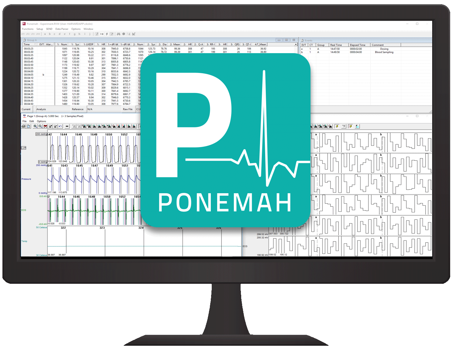
Ponemah CARDIO Software
For over 3 decades Ponemah Software has been trusted by researchers worldwide to discover new insights into their research applications. Ponemah is the data acquisition platform that powers our DSI Implantable Telemetry solutions for cardiovascular research. Ponemah CARDIO software is tailored to acquire, visualize, and report cardiovascular endpoints. A wide range of essential cardiovascular metrics can be derived in real time from the primary signals acquired from in vivo models or ex vivo heart perfusion setups.
- Data acquisition, visualization, and analysis of in vivo or ex vivo cardiovascular hemodynamic and electrophysiologic signals.
- Accurate and consistent results through industry validated algorithms.
- Adjust algorithm settings to optimize signal analysis from various species and unique morphologies.
- Acquisition of primary signals from our DSI* ACQ-7700 signal conditioners or HSE* PLUGSYS Modules as well as third-party analog signals.
- Built-in visualization tools to explore data and gain complete confidence in your results
- Fast-tracks subsequent experiments by saving settings as a reusable Protocol
- Immediate results with real-time data visualization and analysis with visual validation marks.
-
Refine results by re-analyzing data segments post-hoc and/or changing mark placement.
*Data Sciences International, Hugo Sachs Elektronik and Harvard Apparatus are all divisions of Harvard Bioscience, Inc.
To ensure that your system is properly configured as a complete setup that meets your experimental needs, please email us at sales@harvardapparatus.com or call us at 800-597-0580. In Europe, please call +49 7665 92000 or email sales@hugo-sachs.de.
Additional Resources:
- Complete line of Ex Vivo Heart Perfusion Systems
- HSE/HA Tissue Bath Systems
- Isolated Lung or Abdominal Organ Perfusion Systems
- HSE PLUGSYS Cases and Modules
- HSE Data Acquisition Hardware
- DSI ACQ-7700 Cases and Signal Conditioners
- Other Ponemah Platforms including CARDIO, PULMODYN, and BDAS
Data Analysis Modules Included with Ponemah CARDIO*
Each data analysis module derives a wide range of scientifically validated, industry approved cardiovascular endpoints.
- Multi-Lead Electrocardiogram (ECG)
- Blood Pressure (BP)
- Left Ventricular Pressure (LVP)
- Cardiac Volume (CVOL)
- Action Potential (MAP)
- Systemic Blood Flow (SBF)
- Coronary Blood Flow (CBF)
*Expand the specifications section below for a complete list of parameters derived from the primary signals
Reliable Data Services: We also offer Data Analysis as a service. We can assist with the creation of high quality, usable reports that summarize your experimental data and provide you with more time to evaluate the experimental results and plan your future studies. Contact us for more information.
- DSI Scientific Services for assistance in study setup and data analysis.
- Customized report packages for summarizing experimental data.
- Saves time, enhances confidence in results, and facilitates planning for future experiments.
Multi-Lead Electrocardiogram (ECG)
Analyzes ECG signals to provide single and multi-lead calculations. The ECG signal can have positive, negative, or bi-phasic T waves, P waves, and QRS complexes and validation marks, such as for Q, R, S, End of T and Beginning of P are automatically drawn.
Multi-Lead ECG parameters can also be calculated to provide additional information related to QT interval prolongation and dispersion.
The list below describes the parameters calculated either in real-time or during subsequent analysis.
| Name | Definition |
| Num | The number of the cardiac cycle. |
| HR | The heart rate is computed in beats-per-minute. |
| R-H, P-H, T-H | Height of the waves from the Iso-electric level, in millivolts. |
| T-HN | Lowest point between the end of the S wave and the end of the T. |
| ST-I | Time interval in milliseconds from the S wave to end of the following T wave. |
| ST-E | The ST elevation, measured in “ST Measure” milliseconds after the S wave, from the Iso-electric level. |
| QRS | Time interval of the QRS complex. |
| PR-I, QT-I, RR-I | Common Interval measurements. |
| QAT | Q Alpha T is the time interval from the Q wave to the peak of the following T wave. |
| QTcb, QTcf, QTcv, QTcm and QTck | Multiple corrected QT intervals (Bazett, Fridericia, Van de Water, Matsunaga and King). |
| EQTS, EQTSc, EQTM, EQTMcs, EQTMce, QTMc | Various cross channel calculations available with Multilead ECG Analysis. |
| QTD | QT Dispersion available with Multilead ECG Analysis. |
| QR-I, QRSA | QR interval, QR amplitude. |
| MxdV | Maximum derivative of the R wave. |
| T-A | Area of the T wave from the Iso-electric level. |
| PCt, TCt | The number of valid waves encountered in the logging period. |
| QTCt | QT count. |
| Count | Reports the number of ECG cycles in a given logging (averaging) period. |
| BAD | The number of arrhythmic beats detected during a specified logging period. |
| GW, TW | The Good Wave counts and number (Total Wave) of good and bad complexes. |
| QATN | Time, in milliseconds, between the Q wave and the lowest point between the end of S and the end of T wave. |
| PWdth | P Width reports the time, in milliseconds, between the start and end of the P wave. |
| Tpe-I | The time in milliseconds between the peak of the T wave and the end of the T wave. |
| T-P | The signal value at the peak of the T wave relative to the Iso-electric level. |
| Noise | This parameter reports an approximation of the noise level in the ECG cycle. |
Blood Pressure (BP) Analysis
Analyzes arterial and venous pressures. Pulse Wave Velocity calculations are also available when combined with a DSI or HSE hardwired solution using two pressure catheters.
In addition to displaying aortic pressure recordings, validation marks are automatically set for Diastolic, End Diastolic, Systolic. In addition, users can define and display % Recovery Point.
The list below describes the parameters calculated either in real-time or during subsequent analysis.
| Name | Definition |
| Num | The number of the cardiac cycle. |
| Sys, Dia, Mean | Systolic, Diastolic, and Mean pressure. |
| PH, HR, TTPK, ET | Common parameters include Pulse height, Heart rate, Time to peak, Ejection time. |
| +dP/dt, -dP/dt | Maximum positive and negative value of the first derivative of the pressure. |
| %REC | The amount of time it takes the pressure to recover. |
| NPMN | Non-pulsatile mean pressure. |
| Q-A | The Q-A Interval is the time in milliseconds from the start of the Q-wave, in the ECG trigger channel, to the start of the systolic pressure rise. |
| Mean2 | An alternate representation for Mean calculated as (Systolic + 2 * Diastolic)/3. |
| PTT | Pulse Transit Time (PTT) is the time between the prior systolic time of the upstream channel and the systolic time of the selected channel. This time is reported in ms. |
| PWV | Pulse Wave Velocity (PWV) is the velocity calculated by using the Pulse Wave Distance (PWD) and Pulse Transit Time (PTT). PWV is calculated as: Pulse Wave Velocity = Pulse Wave Distance / Pulse Transit Time. |
| IBIs, IBIms, IBIed | Inter-Beat Interval (IBI) is the time in milliseconds between cardiac cycles and allows Heart Rate Variable (HRV) to be calculated from Blood Pressure signals (Frequency Domain). |
| Count | Reports the number of cycles in a given logging period. |
Left Ventricular Pressure (LVP) Analysis
Analyzes pressure signals from the left ventricle and is used as a reference signal for other analysis modules, such as Cardiac Volume. Parameters such as Left Ventricular End Diastolic Pressure, Systolic Pressure, Percent Recovery Times and -dP/dt max can be marked automatically.
The list below describes the parameters calculated either in real-time or during subsequent analysis.
| Name | Definition |
| Num | The number of the cardiac cycle. |
| Sys | The systolic pressure is the maximum pressure that occurs during the cardiac cycle. |
| LVEDP | The left ventricular end diastolic pressure is the pressure at the last zero crossing of the differentiated pressure during the rise to the systolic period. |
| EMw | Electromechanical Window represents the time difference in ms between the end of electrical systole (end of the T wave) and the end of ventricular relaxation. It is a potential biomarker for Torsades de Pointes (TdP) risk that has greater predictability than using QT prolongation. |
| Min | The minimum pressure during the cardiac cycle. |
| TTI | Tension-Time Index is the area under the left ventricular pressure during the ejection phase of the contraction. This is the integration between the LVEDP point and -dP/dtMAX. |
| DP | Developed pressure is the difference between the systolic pressure and the left ventricular end diastolic pressure (SYS-LVEDP). |
| HR | The heart rate is computed in beats-per-minute. It is calculated by taking the reciprocal of the time interval for the cardiac cycle multiplied by 60. |
| +dP/dt | +dP/dt is the maximum positive value of the first derivative of the pressure that occurs during the cardiac cycle. |
| -dP/dt | -dP/dt is the maximum negative value of the first derivative of the pressure that occurs during the cardiac cycle. |
| CI | Contractility index is +dP/dt divided by the pressure at that point. |
| RT1, RT2 | The Relaxation Time is the period from -dP/dt to the time specified by the Relaxation Time attribute. The time is reported in milliseconds. |
| dP (A, B, C, and D) | These parameters report the value of dP/dt at the pressure levels specified in dP/dt A, dP/dt B, dP/dt C, and dP/dt D (in the attributes window). |
| NPMN | The non-pulsatile mean pressure reported for a logging period. This parameter is still reported even if no pulse pressure exists. |
| Q-A | The Q-A Interval is the time in milliseconds from the start of the Q-wave, in the ECG trigger channel, to the start of the systolic pressure rise (LVEDP). |
| IVT | The time in milliseconds from the start of the systolic pressure rise (LVEDP) to the maximum slope of the systolic pressure rise (+dP/dt). |
| TTI-T | LVEDP to -dP/dt. The time is in milliseconds. |
| Tau | Tau is the time constant isovolumic left ventricular pressure decay. It is reported in milliseconds, and can be defined using Pressure, Developed Pressure and dP/dt. |
| Period | The duration of the current cycle time, in milliseconds. |
| Count | Reports the number of cycles in a given logging period. |
Cardiac Volume (CVOL) Analysis
Can analyze any volume from the circulatory system, as measured by a pressure volume catheter, and can derive on a beat-to-beat basis values for the cardiac cycle. Validation marks provide the user with visual documentation that the real-time analysis is providing appropriate cycle recognition.
Additional features built into the Data Review functionality allow the Cardiac Volume channel and LVP channel to be analyzed in a post process mode by the Pressure-Volume Loop analysis. Pressure-Volume Loops permit the display and analysis of left ventricular pressure cycles plotted against cardiac volume cycles.
Pressure-Volume Loop analysis features and calculations:
- Cuvette Calibration - Catheter Selection - Choose from 40 pre-defined PV catheters for volume calibration during acquisition.
- Parallel Volume calculation - Volume component due to parallel conductance. Parallel volume is subtracted from summed volume signal by means of a saline bolus injection or input of empirical data.
- Alpha Correction Factor - Reconcile the volume obtained using conductance technology with the volume obtained by other means. Stroke Volume is divided by Alpha.
- ESPVR (End Systolic PV Relationship) - Slope, X intercept, correlation coefficient.
- EDPVR (End Diastolic PV Relationship) - α, β, X intercept, correlation coefficient.
- PRSW (Pre Recruitable Stroke Work) - Slope, X intercept, correlation coefficient.
The list below describes the parameters calculated either in real-time or during subsequent analysis.
| Name | Definition |
| Num | The number of the cardiac cycle. |
| Vmin | The minimum volume value in a cycle. |
| Vmax | The maximum volume value in a cycle. |
| SV | The stroke volume, which equals Vmax - Vmin. |
| -dPV | The volume at the LVP Min Slope mark. |
| Ped | The pressure at the LVEDP mark. |
| Ved | The volume at the LVEDP mark. |
| %EF | Ejection fraction, calculated as Stroke Volume/Vmax * 100. |
| PFR | Peak filling rate. |
| -dV | Peak emptying rate. |
| TPFR | Time to peak filling rate. |
| DFT | Diastolic Filling Time. |
| %FT | Time to peak filling as a percentage of Diastolic fill time, equals TPFR/DFT * 100. |
| Pes | Pressure at max elastance. |
| Ves | Volume at max elastance. |
| SW | Stroke Work. |
| Ea | Arterial elastance. |
| HR | Heart rate. Computed in beats-per-minute. |
| CO | Cardiac Output. |
Action Potential (MAP) Analysis
Analyzes a host of action potentials with focus on key regions such as plateau, recovery time and upstroke velocity.
The list below describes the parameters calculated either in real-time or during subsequent analysis.
| Name | Definition |
| Num | The number of the cardiac cycle. |
| Max | The Maximum Voltage that occurred during a cardiac cycle. |
| EDV | The voltage prior to the upstroke of the action potential. |
| Min | The minimum voltage that occurred during a cardiac cycle. |
| Plto | The plateau voltage. |
| Amp | PLTO minus EDV. |
| Rate | The beats-per-minute. |
| +dV | The maximum positive rate of change that occurred. |
| -dV | The maximum negative rate of change that occurred. |
| REC1 | The time, in milliseconds, from the beginning of the upstroke of the action potential, to the point where the signal drops below the level corresponding to the % Recovery 1 level. |
| xR1 | The number of cycles that did not reach the % Recovery 1 level, within the logging period. |
| REC2 | The time, in milliseconds, from the beginning of the upstroke of the action potential, to the point where the signal drops below the level corresponding to the % Recovery 2 level. |
| xR2 | The number of cycles that did not reach the % Recovery 2 level, within the logging period. |
| REC3 | The time, in milliseconds, from the beginning of the upstroke of the action potential, to the point where the signal drops below the level corresponding to the % Recovery 3 level. |
| xR3 | The number of cycles that did not reach the % Recovery 3 level, within the logging period. |
| %Max | The time, in milliseconds, from the beginning of the upstroke of the action potential, to the point where the signal recovers to 100% or as close to 100% as possible. |
| Time | The time interval between action potentials. |
| Cnt | The number of cycles in the logging period. |
| RiseT | The time, in milliseconds, between the EDV and Max points. |
Systemic Blood Flow (SBF) Analysis
Analyzes systemic blood flow from the circulatory system and eliminates drift resulting from certain types of flow meters.
The list below describes the parameters calculated either in real-time or during subsequent analysis.
| Name | Definition |
| Num | The number of the cardiac cycle. |
| Mean | The integrated level of the flow for the cardiac cycle. |
| FMax | Maximum Flow that occurred for a cardiac cycle. |
| FMin | Minimum Flow that occurred for a cardiac cycle. |
| CO | Cardiac Output is the mean flow normalized to ml/Min. Note: When running in a logging mode other than 1 epoch, the CO value will be calculated from the averaged mean flow value. |
| SV | Stroke Volume. |
| +dQ | +dQ is the maximum positive value of the first derivative of the flow that occurs during the cardiac cycle. |
| Iso | The value of flow during the rapid up rise of the flow signal. |
| TPR | Total Peripheral Resistance. |
| NPMN | The non-pulsatile mean calculated over the complete logging interval. |
Coronary Blood Flow (CBF) Analysis
Provides coronary flow and volume information during systolic and diastolic periods in conjunction with the LVP module.
The list below describes the parameters calculated either in real-time or during subsequent analysis.
| Name | Definition |
| Num | The number of the cardiac cycle. |
| Mean | The integrated level of the flow for the cardiac cycle. |
| FMax | Maximum Flow that occurred for a cardiac cycle. |
|
FMin |
Minimum Flow that occurred for a cardiac cycle. |
| CO | Cardiac Output. |
| SV | Stroke Volume. |
| +dQ | +dQ is the maximum positive value of the first derivative of the flow that occurs during the cardiac cycle. |
| SFIw | Flow during systolic part of cardiac cycle. |
| DFlw | Flow during diastolic part of cardiac cycle. |
| SVol | Volume during the systolic part of the cardiac cycle. |
| DVol | Volume during the diastolic part of the cardiac cycle. |
| -Flow | Any negative flow that occurred during the cardiac cycle. |
| TVol | Total Volume is the sum of Systolic Volume and Diastolic Volume: SVol + DVol. |
| NPMN | The non-pulsatile mean calculated over the complete logging interval. |

